Macular pucker and macular holes
As we age, the clear gel (called the vitreous) that fills the eye gradually breaks down and becomes more liquid. This process, known as vitreous degeneration, is natural and usually occurs between the ages of 40 and 70. In most cases, it causes no problems. However, sometimes the vitreous pulls too strongly on the retina—the thin, light-sensitive layer at the back of the eye that captures visual images. This pulling, or traction, can lead to conditions such as a macular pucker or a macular hole.
What is a macular pucker?
The macula of your eye is the center of your retina that gives you the super-sharp vision you need to see fine details. When retinal wrinkling occurs in the macula, it’s called “macular pucker.”
If you take a sheet of clear cellophane and stretch it tight before your eyes, you can see right through it. However, if you wrinkle the cellophane and try to see through it, the images become distorted or even impossible to see. This is similar to what happens to your vision when a macular pucker occurs in your eye. Most cases are mild, but some can cause distortion and significant vision loss. Thankfully, this can be corrected.
Read more about Macular Pucker [PDF]What is macular hole?
A macular hole is a tear or hole in the center or your retina which can make your vision blurry and distorted. Recently, effective treatments have been developed to treat macular holes and help restore vision.
Symptoms & The Path to Diagnosis
Macular pucker and macular holes can often start subtly, but recognizing the early signs is key to successful treatment. Both conditions can distort your vision, making it difficult to read, drive, or see faces clearly. Here are some common symptoms to look for:- Wavy or distorted vision: Straight lines, such as doorframes or power lines, may appear bent, wavy, or crooked.
- Blurry central vision: You might notice a blurry or gray spot right in the center of your vision.
- Difficulty reading: It may become challenging to read fine print, even with your glasses.
- Diminished vision: Colors may seem less bright, and your overall vision can become dull.

What to Expect During Your Eye Exam
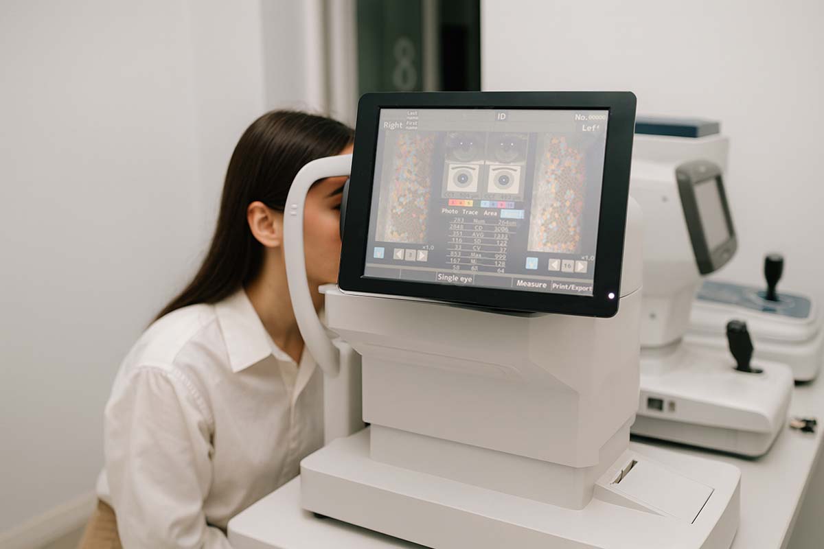
If you experience any of these symptoms, a thorough eye exam is the first step. At Teton Retinal Institute, Dr. Moffett uses the latest diagnostic technology to accurately identify these conditions. During your exam, he may use Optical Coherence Tomography (OCT). This non-invasive imaging device provides a high-resolution, cross-sectional view of your retina, allowing him to see the precise extent of the pucker or hole. This quick and painless scan gives us the detailed information we need to design a personalized treatment plan for you.High-tech treatments provide hope
Today, there are effective treatments for macular holes and macular pucker. In many cases, these new procedures can restore lost vision. Early detection and treatment are the keys to success.
Frequently Asked Questions
-
What's the difference between a macular pucker and a macular hole?
The difference lies in the extent of the damage to the macula. A macular pucker is a wrinkling or scar tissue on the surface of the macula, often described as a "cellophane-like" layer. This wrinkling distorts your central vision, making straight lines appear wavy. A macular hole, on the other hand, is an actual tear or break that forms in the very center of the macula. This break causes a significant blurry spot or blind spot in your central vision, as the light is unable to be focused on that part of the retina.
-
Can macular holes or pucker heal on their own?
Unfortunately, a macular hole will not heal on its own and requires surgery to be repaired. A macular pucker may be mild and not require treatment if it is not significantly affecting vision. However, a pucker that is causing noticeable vision distortion will not resolve on its own and usually requires surgery to remove the scar tissue and restore the retina to its proper shape.
-
Is the surgery painful?
No, the surgery is not painful. The procedure is performed using a local anesthetic, which means the area around your eye will be completely numb. You will be awake but will not feel any pain. The surgeon may also use a sedative to help you feel relaxed and comfortable throughout the procedure. Any post-operative discomfort is typically managed with medication and can be discussed with the doctor.
-
How long does it take to recover from vitrectomy surgery?
The initial recovery from vitrectomy surgery for a macular hole or pucker is typically a few days to a week. Most patients can return to their normal daily activities (with some restrictions) within a week. However, the final vision recovery can be a longer process, sometimes taking several months. This is because the eye needs time to fully heal and for the vision to stabilize. Your doctor will provide you with a specific timeline based on your individual case.
-
Will my vision return to normal after treatment?
The goal of treatment is to restore as much vision as possible and prevent further loss. While many patients experience a significant improvement in their vision after surgery, it's important to have realistic expectations. The degree of vision that returns depends on factors such as how long the condition was present before treatment. For many patients, the surgery is very successful in improving their vision, reducing distortion, and preserving their sight for the future.

Protect Your Vision with Expert Retinal Care
If you’re experiencing blurry or distorted central vision, don’t wait. Early evaluation and treatment can help preserve your eyesight and improve your quality of life. Our retina specialists use advanced imaging and treatment options to provide the best possible outcomes.
Schedule Your Retinal Evaluation or give us a call at 208-535-4900